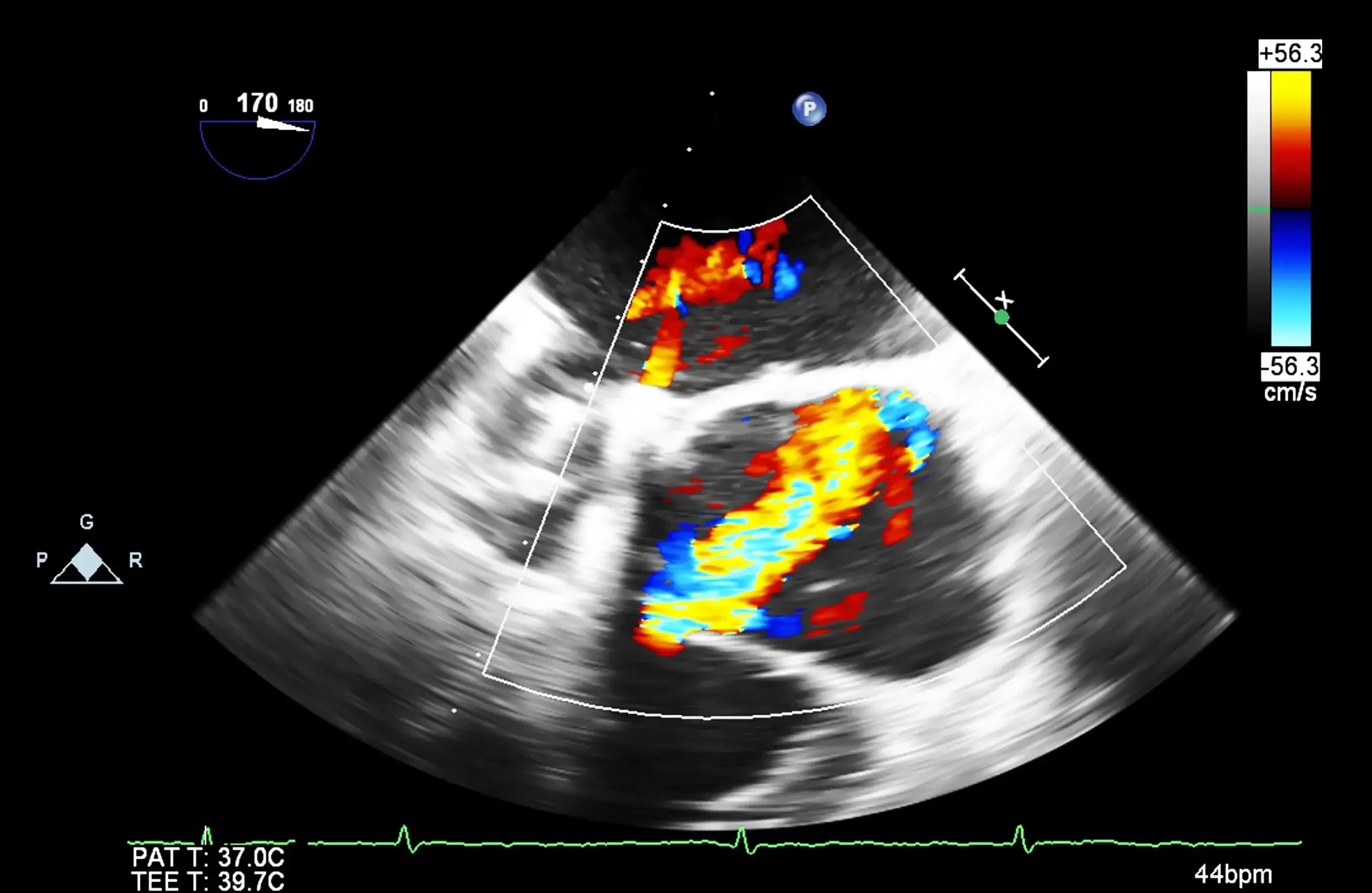
2D and 3D Echocardiography
At Apex Heart Hospital, we use the latest advancements in cardiac imaging to provide accurate diagnoses and personalized treatment plans. 2D and 3D echocardiography are among the most essential, non-invasive tools in our diagnostic arsenal. These ultrasound-based imaging techniques help our cardiologists visualize the heart in real-time, assess its function, and detect a wide range of heart diseases.
What is Echocardiography?
Echocardiography (also called an “echo”) is a safe, painless test that uses high-frequency sound waves (ultrasound) to create images of the heart. It shows the heart’s chambers, valves, walls, and blood vessels, and evaluates how effectively the heart is pumping blood.
2D Echocardiography: The Standard Diagnostic Tool
2D echocardiography provides flat, cross-sectional images of the heart. This is the most commonly used form of echocardiography and is essential for evaluating:
- Heart size and structure
- Pumping function (ejection fraction)
- Valve movement and leakage (regurgitation or stenosis)
- Pericardial effusion (fluid around the heart)
- Congenital heart defects
- Wall motion abnormalities post-heart attack
- During the test, a handheld device called a transducer is moved over the chest, and images appear on a monitor in real-time. The test typically takes 15 to 30 minutes and requires no special preparation.
3D Echocardiography: Advanced Heart Imaging
3D echocardiography builds upon 2D imaging by creating three-dimensional, real-time visualizations of the heart. It provides a more complete and accurate picture of the heart’s anatomy and function. This is particularly helpful in:
- Pre-surgical planning for valve repair or replacement
- Detailed valve assessment (mitral, aortic, etc.)
- Diagnosing complex congenital heart defects
- Evaluating left ventricular volume and function with greater precision
- Guiding catheter-based interventions such as TAVR or device closures
- At Apex Heart Hospital, we use high-end echocardiography systems capable of real-time 3D imaging, offering unmatched detail and diagnostic confidence.
Specialized Types of Echocardiography Offered
In addition to standard transthoracic echo (TTE), we offer specialized techniques:
1. Transesophageal Echocardiography (TEE)
An ultrasound probe is inserted into the esophagus for clearer images of the heart’s back structures, often used in surgical cases or stroke evaluation.
2. Stress Echocardiography
Combines echo imaging with physical or drug-induced stress to assess how the heart performs under exertion—especially useful in detecting coronary artery disease.
See Your Heart Like Never Before
- Whether for routine heart screening or detailed evaluation of a complex condition, 2D and 3D echocardiography at Apex Heart Hospital provides clear, real-time insights into your heart’s health. Our commitment to precision diagnosis ensures that every patient receives the best care, from detection to recovery.
Take the first step toward a healthier heart — book your echo test with us today.


